Faculty
Dr. Jayadevan E. R.
Professor & HeadDr. C. Kesavadas
Professor Senior GradeDr. Bejoy Thomas
Professor Senior GradeDr. Anoop A.
Additional ProfessorDr. Jineesh V
Additional ProfessorDr. Ajay Alex
Associate ProfessorFacilities
256 SLICE CT SCANER (Philips Brilliance 256 slice)
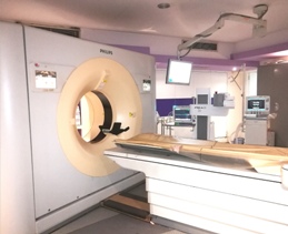
The multi slice CT has revolutionized cross sectional imaging with exquisitely detailed multiplanar depiction of human anatomy and functional details. 256-slice is one of the most advanced CT systems in the world. It delivers high image quality, dose efficiency and rapid reconstruction times and can carryout a full body scan in less than a minute.
Features and applications
DUAL SLICE CT Scanner (GE)
Dual slice (multislice) CT provides speed, resolution, image quality, reduced dose, and coverage optimal for an emergency CT study. Sub millimeter slice thickness provides the high spatial resolution required for the visualization of small anatomy. Speed is sufficient to perform more aggressive studies like CT angiograms.
MAGNETIC RESONANCE IMAGING 3 TESLA (GE Discovery 750w)
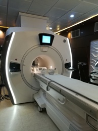
One of the leading mri system with wide bore and silent scan technology to benefit the patients for a comfortable scanning experience.capability of high definition structural and functional imaging in both neuro and cardiovascular applications provides superlative clinical diagnostic benefits.
Features and applications
MAGNETIC RESONANCE IMAGING (Siements Avanto 1.5T)
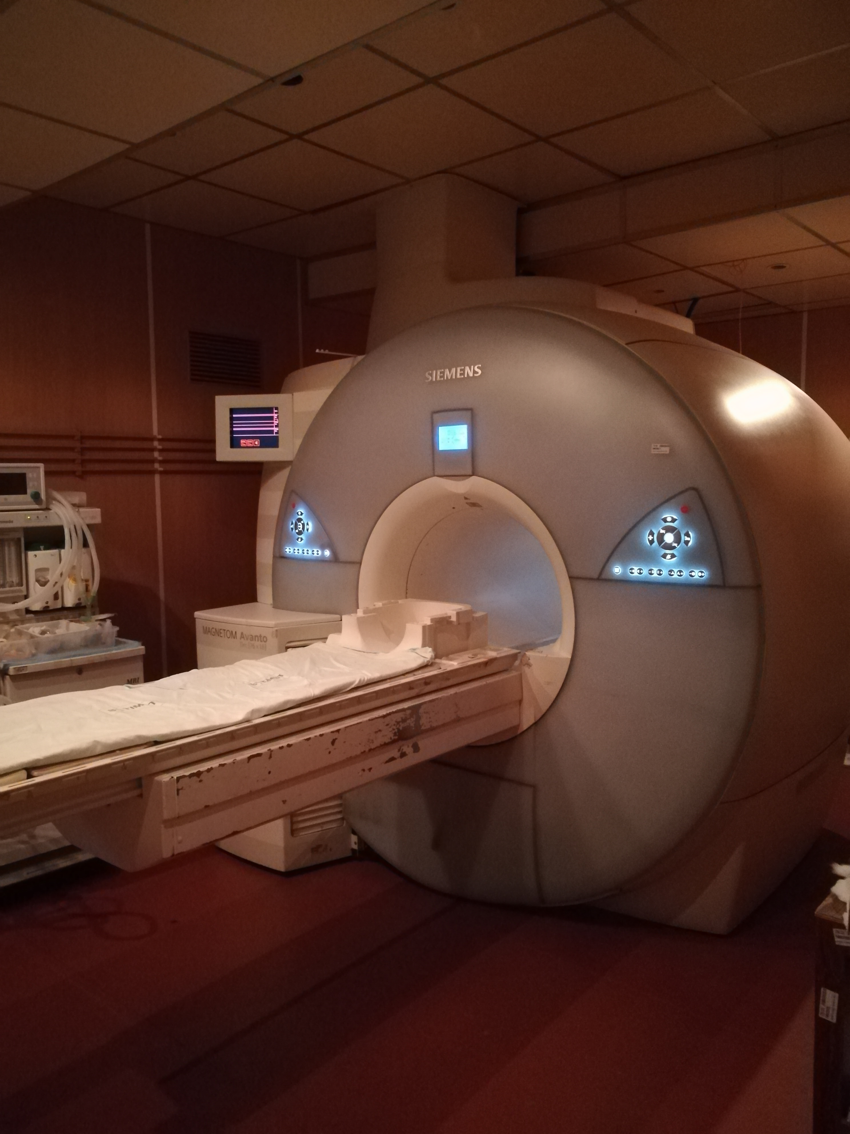
High end award winning 1.5T MR system, capable of performing one of the most comprehensive applications ranges available today. The unique combination of leading magnet and gradient technology together with revolutionary image acquisition techniques provides unsurpassed clinical benefits. Excellent and efficient parallel imaging technique enhances the flexibility, accuracy and speed of imaging examinations.
Features and applications
NEURO INTERVENTION CENTRE
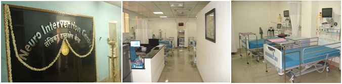
Institute has started Neuro Intervention Centre (NIC) in January 2013, comprising of dedicated neuro intensive care unit, general ward and state of the art Cath lab. Neuro intervention centre (NIC) is a tertiary care facility for the comprehensive management of patients suffering from various neuro-vascular disorders. The quality management practices coupled with a strong multidisciplinary co-operative directions of NIC has contributed significantly in achieving less than 1% morbidity and mortality in the neurovascular treatment. This facility is the only one of this kind in India regarding the management of neurovascular diseases.
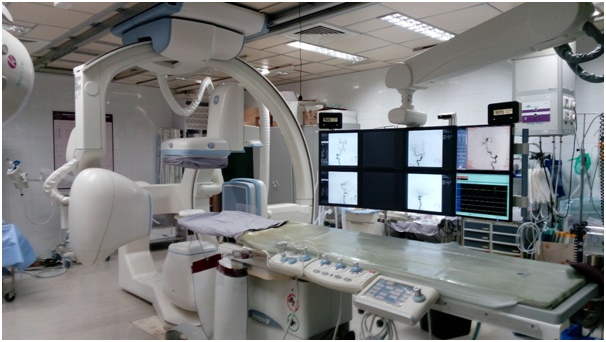
We have started second cath lab in 2017 (Philips AlluraXper FD 20 Single plane).This is mainly to manage huge need of diagnostic neuro angiographic procedures and other vascular and non-vascular diseases.
Interventional radiology division is managing more than 4000 patients in the outpatient section and performing more than 1200 inpatient procedures per year at present (data as on 2017).
Services
Cath labs
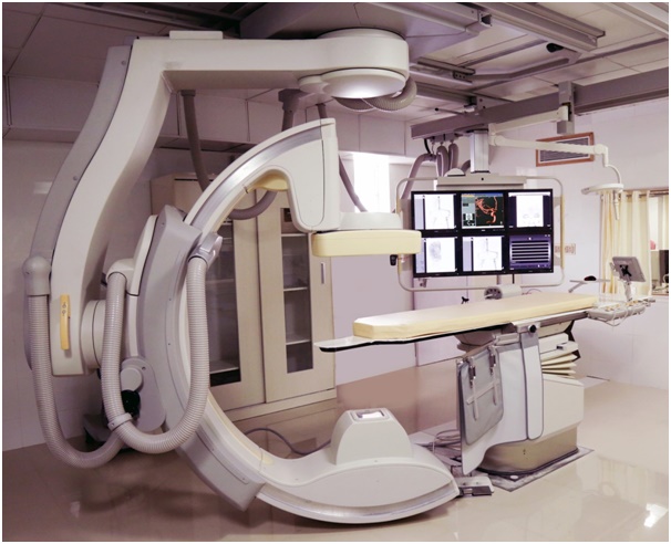
DIGITAL SUBTRACTION ANGIOGRAPHY (GE Innova 4100 biplane)
The DSA system equipped with biplane detectors give breathtaking fluoroscopy and outstanding angiography and is one of the most advanced DSA systems in the world. This dedicated neuro vascular DSA system with large 40 x 40cms size flat panel tremendously decrease procedure time and radiation to the patient.
The superb image quality and advanced 3D tools help in confident decision making during all kinds of endovascular procedures. High resolution imaging by the fixed X-ray system provides crisp, virtually distortion-free visualization of small details and objects to support endovascular interventions, including intracranial aneurysm coiling and carotid stenting procedures.
Features and applications
Ultrasound system - Philips iU22
The premium performance ultrasound system captures crisp, high-resolution images even in technically challenging situations. It makes easy to add 3D imaging to any exam by removing the barriers to volume imaging and a host of workflow enhancers facilitate faster exams, enabling perfect image optimization.
Features
PACS (Picture archiving and communication system)
Makes the department more efficient, enables increased productivity, and allows department to more effectively serve hospital staff, patients, and referring physicians. It forms a comprehensive, fully-integrated solution that meets all digital needs. In addition to RIS, the system features state of the art Picture Archiving and Communication System (PACS), medical reporting, solution monitoring & management services (SMMS), and audit services. Integrated with other hospital systems, such as hospital information systems (HIS) and Electronic Patient Records (EPR), the benefits of RIS extend well beyond radiology.
Features
Radiofrequency Ablation - Celon POWER
Radiofrequency ablation system provides the latest innovative radiofrequency technology for the precise and efficient ablation of tumors, varicose veins, inter vertebral disc diseases, denervation and other diseases using bipolar radiofrequency. Main benefit of the bipolar technology is the restriction of radiofrequency current flow to the volume under treatment (e.g. tumour), as a result of which undesirable side-effects are avoided. In addition to the above, no neutral electrodes are required, which means simplification of the patient's preparation, cost reduction and increased safety.
Features
Computed and Digital Radiography (CR and DR) - Agfa and Cura
Flexible, high quality digital imaging solution enables seamless integration of the X-ray generator with the hospital or radiology information system. Innovative high end advanced flat panel detectors provide outstanding image quality, optimal workflow and operability.
Features
Mentors
Prof. Mahadevan Pillai was born in Kayamkulam (Kerala) in 1908. He took his medical training at Vizag Medical college. After completing a Diploma in Medical Radiology from UK, he worked in the Dept of Radiology at Madras Medical College (Barnard Institute). It is here that he popularized the use of neuroradiological investigations, in association with Dr.Ramamurthi. After short stints as professor of radiology at NIMHANS and Medical College Trivandrum, he joined the institute as the head of the department of radiology. He retained the position from 1975 to 1979. During his tenure at this department, he set high standards of quality for radiological investigations.
Prof.Sashidharan who graduated from Banaras Hindu University and also completed his postgraduate training in radiology at the same institute and joined the institute as an assistant professor in 1976. He was later promoted to the post of associate professor and then headed the department from 1981 - 1983.
Prof.V.R.K.Rao (Vedula Rajani Kanth Rao) was born at Anakapalle in Andhra Pradesh in 1948. He graduated from Andhra Medical College, Visakhapatnam in 1970 and pursued his postgraduate program in Radiology at Safdarjang Hospital, Delhi University and at Banaras Hindu University. He joined the department as Assistant Professor in Radiology in early 1977 and later was in charge of the department from 1986 till 1993 as Professor and Head. He contributed to the birth and growth of Interventional Radiology at this Institute with recognition of the Institute at the national level. Simultaneously useful efforts were made by him to develop biomaterials for Therapeutic Endovascular Neurointervention.
Prof. Kolathu Ravi Mandalam was born in New Delhi in 1954. He graduated from Maulana Azad Medical College, New Delhi and completed his post graduation in radiology from the same institute. After a short period of residency at Safdarjung Hospital, New Delhi, he joined the Department of radiology at SCTIMST. As a team with Prof.V.R.K. Rao and later as the head of the department, he has guided the department to national fame in the field of diagnostic and interventional cardiovascular radiology.
Prof. Arun Kumar Gupta
Prof. Santhosh Joseph
Prof. Kapilamoorthy TR
Services
The department of Imaging Sciences & interventional Radiology as the name suggests, caters to all the diagnostic & interventional needs of the institute. The department has - Xray machines. X-ray of all body parts as needed can be taken. There are also 4 mobile units which are used to take x-rays of patients who cannot be moved to the x-ray unit. For these patients, X-ray can be taken in the bed itself.
ULTRASOUND/ COLOUR DOPPLER:
Ultrasound scanning for all body parts can be carried out at the department when necessary. Doppler imaging can be carried out for the neck & peripheral arteries & veins as also for the visceral vessels. Transcranial arterial doppler can also be done. Ultrasound guided intervnetional procedures performed in the department include aspiration, biopsies, pseudoaneurysm occlusion to name a few.
CT:
With the state of art CT scanner, the department caters to all types of CT studies such as non contrast & contrast studies of the brain, abdomen, lung. Cardiac CT, coronary CT, CT angiography for the vessels of the brain, abdomen, lung and peripheral arteries can also be performed.
Entire range of CT guided interventional procedures like aspiration / biopsy and therapeutic procedures like CT guided drainage procedures, ablation of tumors, CT guided alcohol injection into vertebral tumors, CT guided laser ablation of osteoid osteoma ( a bone tumor ) etc are performed.
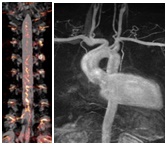
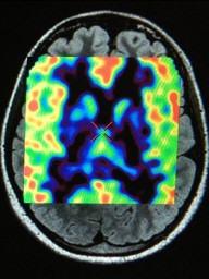
MRI:
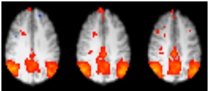
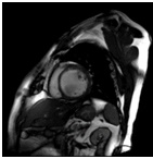
MR offers a vide range of diagnostic options. This includes routine imaging as also advanced imaging protocols. These include Cardiac MRI, MR angiography, diffusion weighted imaging, perfusion weighted imaging, MR spectroscopy, Diffusion tensor imaging, functional MRI.
DIGITAL SUBSTRACTION ANGIOGRAHY UNIT:
The department offers services for a wide variety of vascular lesions - intracranial as also extracranial. This includes peripheral arterial angioplasty, intracranial aneurysm coiling, intracranial arteriovenous malformation embolisation, embolisation of spinal vascular malformations, tumour embolisation, vertebroplasty to name a few. A detailed list of these procedures can be obtained from the website.
Neuro endovascular and Interventional Radiological Procedures
Endovascular Angioplasty (PTA & STENTING)
Endovascular Thrombolysis
Endovascular Embolization
Aneurysms
AVMs
VOGM
Non Galenic AV Fistulas
Dural arteriovenous fistula (DAVF)
Carotid-cavernous fistula (CCF)
Spinal vascular malformation.
Craniofacial vascular malformation.
HNF Tumours
Spinal tumours.
Epistaxis
Post traumatic arteriovenous fistula
Traumatic arterial Injuries
Endovascular Treatment of Vasospasm
Papavarine infusion
Balloon Angioplasty
Endovascular Blood sampling from
Inferior Petrosal sinus and cavernous sinus
Miscellaneous endovascular Procedure
Superselective Chemotherapy of brain tumours
Temporary balloon occlusion test of the internal carotid artery
Wada's test
Direct percutaneous glue injection in tumours.
Peripheral Endovascular and Intervention Radiological Procedures
Arterial Endovascular Percutaneous Tranluminal non-coronary angioplasty and /or stenting of
Endovascular Thrombolysis
Peripheral vascular occlusion
Acute thromboembolism
Vascular graft occlusion
Endovascular Embolization
Peripheral aneurysm embolization
Peripheral AVM embolization
Pulmonary AVM/AVF embolization
Hemoptysis
Gastro intestinal bleeding
Epistaxis
Uterine artery embolization
Testicular vein embolization vericoceles
Ovarian Vein embolization
Acute haematuria due to disease process or post nephrostomy
Aortopulmonary collaterals before or after surgery for congenital heart disease
Endovascular Peripheral AVF Closure
Stent Graft placements
Endovascular Venous Procedures
Superior Vena caval venoplasty and stenting
Inferior Vena cava venoplasty and stenting
Inferior Vena cava filter placement for deep vein thrombosis
Varicose vein ablation using laser or radio frequency.
Non- Vascular Intervention
Musculoskeletal procedure and Backache Management
Percutaneous vertebroplasty
Steroid injection for painful facet joint
Epidural injection of steroid / xylocaine.
Laser Assisted nonvascular intervention
Percutaneous laser disc decompression.
Laser ablation of osteoid osteoma.
Trachea Bronchial Procedures
Tracheal stenting
Laser ablation of bronchial tumours.
Biopsy of lung, mediastinum and chest wall
Miscellaneous Procedures
Laser of RF ablation of tumours of liver, bone, lung and other organs.
Abscess drainage
Retrieval of foreign bodies
Biopsy of abdominal lesion
CT/US guided aspiration
CT/US guided needle biopsy
Emergency Procedures
Thrombolysis of Acute stroke syndrome
Acute tumour bleeding
Acute epistaxis
Intracranial Aneurysmal Bleed
Treatment of cerebral Vasospasm
Angioplasty of vasospasm
Posttraumatic facial or nasal bleeding
Carotid and vertebral injury embolization
Acute Gastrointestinal bleeding
Embolization of acute hematuria
Embolization of Primary or secondary Postpartum bleeding
Embolization Acute bleeding from gynecological Malignancy
Embolization Acute Mesentric Ischemia
Embolization Acute bleeding from Nasopharyngeal Angiofibroma
Embolization of Aortopulmonary collaterals in emergency for congenital heart disease
Ongoing Projects
- Biochemical and functional investigation of dorsolateral prefrontal cortex in mild cognitive impairment using functional magnetic resonance spectroscopy and functional magnetic resonance imaging.
- A resting state fMRI and task based fMRI study: Optimization, language lateralization, memory lateralization and connectivity in normal subjects versus patients with epilepsy.
- Hypoxia and Mineralisation in Alzheimer's disease Detected in vivo with Magnetic Resonance Imaging.
- Brain computer interface & Functional near infra red spectroscopy in stroke rehabilitation.
- Development of tool to prevent contrast extravasation during intravenous injection.
- Virtual reality-based solution for effective neuroanatomy teaching.
- 3D printing for paediatric congenital heart diseases.
- Development of novel prototype mechanical clot retriever for the treatment of acute cerebral ischemic stroke.
- Development of a prototype Flow Diversion Intracranial Stent for the treatment of Complex Intracranial Aneurysms.
- Radiopaque liquid embolization device by chemical grafting of Iodinated compounds onto the ethylene vinyl alcohol co-polymer.
Publications
2017
- Mohimen A, Kumar Kannath S, Jayadevan ER. Skull Base Osseous Arteriovenous Fistula-A Rare Clinical Entity: Case Report and Literature Review.World Neurosurg. 2017 Jan;97:760.
- Arun KM, Smitha KA, Rajesh PG, Kesavadas C. Functional near-infrared spectroscopy is in moderate accordance with functional MRI in determining lateralisation of frontal language areas. Neuroradiol J. 2017 Jan 1.
- Mukherjee A, Muthusami P, Mohimen A, K S, B B, Pn S, Kesavadas C. Noncontrast Computed Tomography versus Computed Tomography Angiography Source Images for Predicting Final Infarct Size in Anterior Circulation Acute Ischemic Stroke: a Prospective Cohort Study.J Stroke Cerebrovasc Dis. 2017 Feb;26(2):339-346.
- Kannath SK, Thomas B, SankaraSarma P, EnakshyRajan J. Impact of non-contrast enhanced volumetric MRI-based feeder localization in the treatment of spinal duralarteriovenous fistula.J Neurointerv Surg. 2017 Feb;9(2):178-182.
- Nagesh C, Asranna A, K P D, Cherian A, Nanda S, Thomas B. Culpable brain lesion causing complex partial status in Wilson's disease: Deduction by arterial spin labeled perfusion MRI.Seizure. 2017 Mar;46:50-52.
- Swaika S, Kannath SK, Rajan JE. Intraprocedural rupture during catheter angiogram in a case of aggressive duralarteriovenous fistula.Neurol India. 2017 Mar-Apr;65(2):378-379.
- Hiremath SB, Muraleedharan A, Kumar S, Nagesh C, Kesavadas C, Abraham M, Kapilamoorthy TR, Thomas B. Combining Diffusion Tensor Metrics and DSC Perfusion Imaging: Can It Improve the Diagnostic Accuracy in Differentiating Tumefactive Demyelination from High-Grade Glioma?AJNR Am J Neuroradiol. 2017 Apr;38(4):685-690.
- Jain NK, Kannath SK, Kapilamoorthy TR, Thomas B. The application of susceptibility-weighted MRI in pre-interventional evaluation of intracranial duralarteriovenous fistulas.J Neurointerv Surg. 2017 May;9(5):502-507.
- Smitha K A, Arun K M, Rajesh P G, Bejoy Thomas, C Kesavadas. Resting state seed based analysis: an alternative to task based language fMRI and its laterality Index. AJNR Am J Neuroradiol. 2017 Jun;38(6):1187-1192.
- Mishra A, Thomas B, Kapilamoorthy TR. Susceptibility weighted imaging - a problem-solving tool in differentiation of cerebellopontine angle schwannomas and meningiomas.Neuroradiol J. 2017 Jun;30(3):253-258.
- Kannath SK, Rajan JE, Mukherjee A, Sarma P S. Factors Predicting Spontaneous Thrombosis of Aggressive Cranial Dural Arteriovenous Fistulas.World Neurosurg. 2017 Jul;103:821-828.
- Smitha KA, Akhil Raja K, Arun KM, Rajesh PG, Thomas B, Kapilamoorthy TR, Kesavadas C. Resting state fMRI: A review on methods in resting state connectivity analysis and resting state networks.Neuroradiol J. 2017 Aug;30(4):305-317.
- Kumar S, Kesavadas C, Thomas B. Susceptibility-weighted Imaging Torch Fire Sign in a Patient with Dystonia due to Hypoxic-ischemic Injury.Ann Indian Acad Neurol. 2017 Jul-Sep;20(3):319.
- Gopinath M, Nagesh C, Santhosh K, Jayadevan ER. Dementia and Parkinsonism-a Rare Presentation of Intracranial Dural Arteriovenous Fistulae.Neurointervention. 2017 Sep;12(2):125-129.
- Kumar S, Nagesh CP, Thomas B, Radhakrishnan A, Menon RN, Kesavadas C. Arterial spin labeling hyperperfusion in Rasmussen's encephalitis: Is it due to focal brain inflammation or a postictal phenomenon?J Neuroradiol.2017 Sep 18.
- Pillai SH, Raghavan S, Mathew M, Gopalan GM, Kesavadas C, Sarma S, Thomas SV. Juvenile Myoclonic Epilepsy with Frontal Executive Dysfunction is Associated with Reduced Gray Matter Volume by Voxel-based Morphometry.Ann Indian Acad Neurol. 2017 Jul-Sep;20(3):270-273.
- Kannath SK, Rajan JE, Sarma SP. Anatomical localization of the cavernous sinus dural fistula by 3D rotational angiography with emphasis on clinical and therapeutic implications.J Neuroradiol. 2017 Sep;44(5):326-332.
- Sheelakumari R, VenkateswaranRajagopalan, Chandran A, Varghese T, Zhang L, Yue GH, Mathuranath PS, Kesavadas C. Quantitative analysis of grey matter degeneration in FTD patients using fractal dimension analysis.Brain Imaging Behav. 2017 Oct 30.
- Kannath SK, Malik V, Rajan JE. Isolated Subcallosal Artery Infarction Secondary to Localized Cerebral Vasospasm of Anterior Communicating Artery Complex Following Subarachnoid Hemorrhage.WorldNeurosurg. 2017 Nov;107:1043.
- Nagesh CP, Mohimen A, Kannath SK, Rajan JE. Primary intraventricularhaemorrhage due to rupture of giant varix of the basal vein of Rosenthal in a patient with long-standing direct CCF: angiographic features and treatment considerations.BMJ Case Rep. 2017 Nov 16;2017.
- Kannath SK, Rajan JE, N SP, P SS, Sukumaran S, Sreedharan SE, Kapilamoorthy TR. Dwell Time Of Stentriever Influences Complete Revascularisation And First Pass Tici3 Revascularisation In Acute Large Vessel Occlusive Stroke.World Neurosurg. 2017 Nov 4.
- Harsha KJ, Jagtap SA, Kapilamoorthy TR, Kesavadas C, Thomas B, Radhakrishnan N. CNS small vessel vasculitis: Distinct MRI features and histopathological correlation. Neurol India. 2017 Nov-Dec;65(6)
2016
- Mohimen A, Kumar K S, Jayadevan ER, Jain NK, Kapilamoorthy TR. Spinal venous hypertension secondary to pelvic extra-spinal arteriovenous fistula-a previously unreported cause of congestive myelopathy.Spine J. 2016 Feb;16(2):e41-2.
- Sheelakumari R, M. Madhusoodanan, A. Radhakrishnan, G. Ranjith, and B. Thomas. A Potential Biomarker in Amyotrophic Lateral Sclerosis: Can Assessment of Brain Iron Deposition with SWI and Corticospinal Tract Degeneration with DTI help?.AJNR Am J Neuroradiol. 2016 Feb; 37(2):252-8.
- Swaika S, Thomas B, Kapilamoorthy TR. Solitary Infantile Myofibroma of Left Ethmoid Sinus With Intracranial Extension.Pediatr Neurol. 2016 Apr;57:107-8.
- Kannath SK, Alampath P, EnakshyRajan J, Thomas B, SankaraSarma P, Tirur Raman K. Utility of 3D SPACE T2-weighted volumetric sequence in the localization of spinal duralarteriovenous fistula.J Neurosurg Spine. 2016 Jul;25(1):125-32.
- Srinivasan K, Thomas B, Shah D, Kannath SK, Menon G, Sandhyamani S, Kesavadas C, Kapilamoorthy TR. Quantification of diffusion and anisotropy in intracranial epidermoids using diffusion tensor metrics and p: q tensor decomposition.J Neuroradiol. 2016 Dec;43(6):363-370.
2015
- James JS, Kumari SR, Sreedharan RM, Thomas B, Radhkrishnan A, Kesavadas C. Analyzing functional, structural, and anatomical correlation of hemispheric language lateralization in healthy subjects using functional MRI, diffusion tensor imaging, and voxel-based morphometry.Neurol India. 2015 Jan-Feb;63(1):49-57.
- James JS, Radhakrishnan A, Thomas B, Madhusoodanan M, Kesavadas C, Abraham M, Menon R, Rathore C, Vilanilam G. Diffusion tensor imaging tractography of Meyer's loop in planning resective surgery for drug-resistant temporal lobe epilepsy.Epilepsy Res. 2015 Feb;110:95-104.
- Srinivasan K, Thomas B. Teaching neuroimages: optic nerve glioma with perineural arachnoid gliomatosis in a patient with neurofibromatosis-1.Neurology. 2015 Mar 31;84(13).
- Chandrasekhar Pammi VS, PillaiGeethabhavan Rajesh P, Kesavadas C, Rappai Mary P, Seema S, Radhakrishnan A, Sitaram R. Neural loss aversion differences between depression patients and healthy individuals: A functional MRI investigation.Neuroradiol J. 2015 Apr;28(2):97-105.
- Jain NK, Swaika S, Thomas B, Kesavadas C, Kapilamoorthy TR. Varying clinical and imaging outcomes in patients with spontaneous thrombosis of vein of Galen malformation--a report of two cases.Childs Nerv Syst. 2015 May;31(5):809-13.
- Mathur A, Jain N, Kesavadas C, Thomas B, Kapilamoorthy TR. Imaging of skull base pathologies: Role of advanced magnetic resonance imaging techniques.Neuroradiol J. 2015 Aug;28(4):426-37.
- Smitha KA, Gupta AK, Jayasree RS. Relative percentage signal intensity recovery of perfusion metrics-an efficient tool for differentiating grades of glioma. Br J Radiol. 2015 Aug;88(1052):20140784.
- Smitha KA, Gupta AK, Jayasree RS. Fractal analysis: fractal dimension and lacunarity from MR images for differentiating the grades of glioma. Phys Med Biol. 2015 Sep 7;60(17):6937-47.
2014
- James JS, Rajesh P, Chandran AV, Kesavadas C. fMRI paradigm designing and post-processing tools.Indian J Radiol Imaging. 2014 Jan;24(1):13-21.
- Hingwala D, Thomas B, Radhakrishnan A, Suresh Nair N, Kesavadas C. Correlation between anatomic landmarks and fMRI in detection of the sensorimotor cortex in patients with structural lesions.ActaRadiol. 2014 Feb;55(1):107-13.
- Muthusami P, Kesavadas C, Kapilamoorthy TR. Radiologic technologists' radiation safety knowledge in India: a multicenter survey.Radiol Technol. 2014 Mar-Apr;85(4):360-8.
- Muthusami P, James J, Thomas B, Kapilamoorthy TR, Kesavadas C.Diffusion tensor imaging and tractography of the human language pathways: moving into the clinical realm.J MagnReson Imaging. 2014 Nov;40(5):1041-53.
2013
- Hingwala D, Kesavadas C, Sylaja PN, Thomas B, Kapilamoorthy TR. Multimodality imaging of carotid atherosclerotic plaque: Going beyond stenosis.Indian J Radiol Imaging. 2013 Jan;23(1):26-34.
- Kesavadas C. Resting state functional magnetic resonance imaging: an emerging clinical tool.Neurol India. 2013 Mar-Apr;61(2):103-4.
- Hingwala DR, Kesavadas C, Thomas B, Kapilamoorthy TR, Sarma PS. Imaging signs in idiopathic intracranial hypertension: Are these signs seen in secondary intracranial hypertension too?Ann Indian Acad Neurol. 2013 Apr;16(2):229-33.
- Hingwala DR, Radhakrishnan N, Kesavadas C, Thomas B, Kapilamoorthy TR, Radhakrishnan VV. Neuroenteric cysts of the brain-comprehensive magnetic resonance imaging.Indian J Radiol Imaging. 2013 Apr;23(2):155-63.
- Santhosh K, Jayadevan ER, Kapilamoorthy TR, Sylaja PN. Temporal evolution of hypertrophied vasa vasorum of common carotid artery triggered by cerebral ischemia: a serial angiographic investigation.Neurol India. 2013 May-Jun;61(3):312-4.
- Hingwala DR, Kesavadas C, Thomas B, Kapilamoorthy TR. Susceptibility weighted imaging in the evaluation of movement disorders.ClinRadiol. 2013 Jun;68(6).
- Muthusami P, Kesavadas C, Sylaja PN, Thomas B, Harsha KJ, Kapilamoorthy TR. Implicating the long styloid process in cervical carotid artery dissection. Neuroradiology. 2013 Jul;55(7):861-7.
- Smitha KA, Gupta AK, Jayasree RS. Total magnitude of diffusion tensor imaging as an effective tool for the differentiation of glioma. Eur. J. Radiol. 2013;82(5):857-861.
- Harsha KJ, Basti RS, Kesavadas C, Thomas B. Susceptibility-weighted imaging in carotido-cavernous fistulas. A case control study.IntervNeuroradiol. 2013 Dec;19(4):438-44.
Peer reviewed journals
- Smitha KA, Gupta AK, Jayasree RS. Total magnitude of diffusion tensor imaging as an effective tool for the differentiation of glioma. Eur. J. Radiol. 2013;82(5):857-861. Impact factor: 2.462.
- Smitha KA, Gupta AK, Jayasree RS. Relative percentage signal intensity recovery of perfusion metrics-an efficient tool for differentiating grades of glioma. Br J Radiol. 2015 Aug;88(1052):20140784. Impact factor: 2.050.
- Smitha KA, Gupta AK, Jayasree RS. Fractal analysis: fractal dimension and lacunarity from MR images for differentiating the grades of glioma. Phys Med Biol. 2015 Sep 7;60(17):6937-47.Impact factor:2.761.
- Jiji S, Smitha K A, Gupta A K, MahadevanPillai V P, Jayasree R S. Segmentation and volumetric analysis of the caudate nucleus in Alzheimer's disease Eur. J. Radiol.2013 ;82:1525-1530. Impact factor: 2.462.
- Smitha K A, Arun K M, Rajesh P G, Bejoy Thomas, C Kesavadas. Resting state seed based analysis: an alternative to task based language fMRI and its laterality Index. AJNR Am J Neuroradiol. 2017 Jun;38(6):1187-1192.Impact factor: 3.67.
- K A Smitha, Akhil Raja, K M Arun, P G Rajesh, Bejoy Thomas, Kapilamoorthy, C Kesavadas. Resting state fMRI: A review on methods in resting state connectivity analysis and resting state networks; Neuroradiol J. 2017 .Aug;30(4):305-317.pubmed indexed.
- Arun K M, Smitha K A, Rajesh P G, C Kesavadas. Functional near infrared spectroscopy is in moderate accordance with functional MRI in determining lateralization of the frontal language areas. Neuroradiol J. 2017. pubmed indexed.